Home » Products » Scanning Electron Microscopy » Focused Ion Beam-Scanning Electron Microscopes (FIB-SEM) » Tescan FIB-SEM for Life Science
Tescan FIB-SEM for Life Science
|
High-Resolution Imaging and 3D Structural Analysis of Biological Samples
Tescan FIB-SEM for Life Science
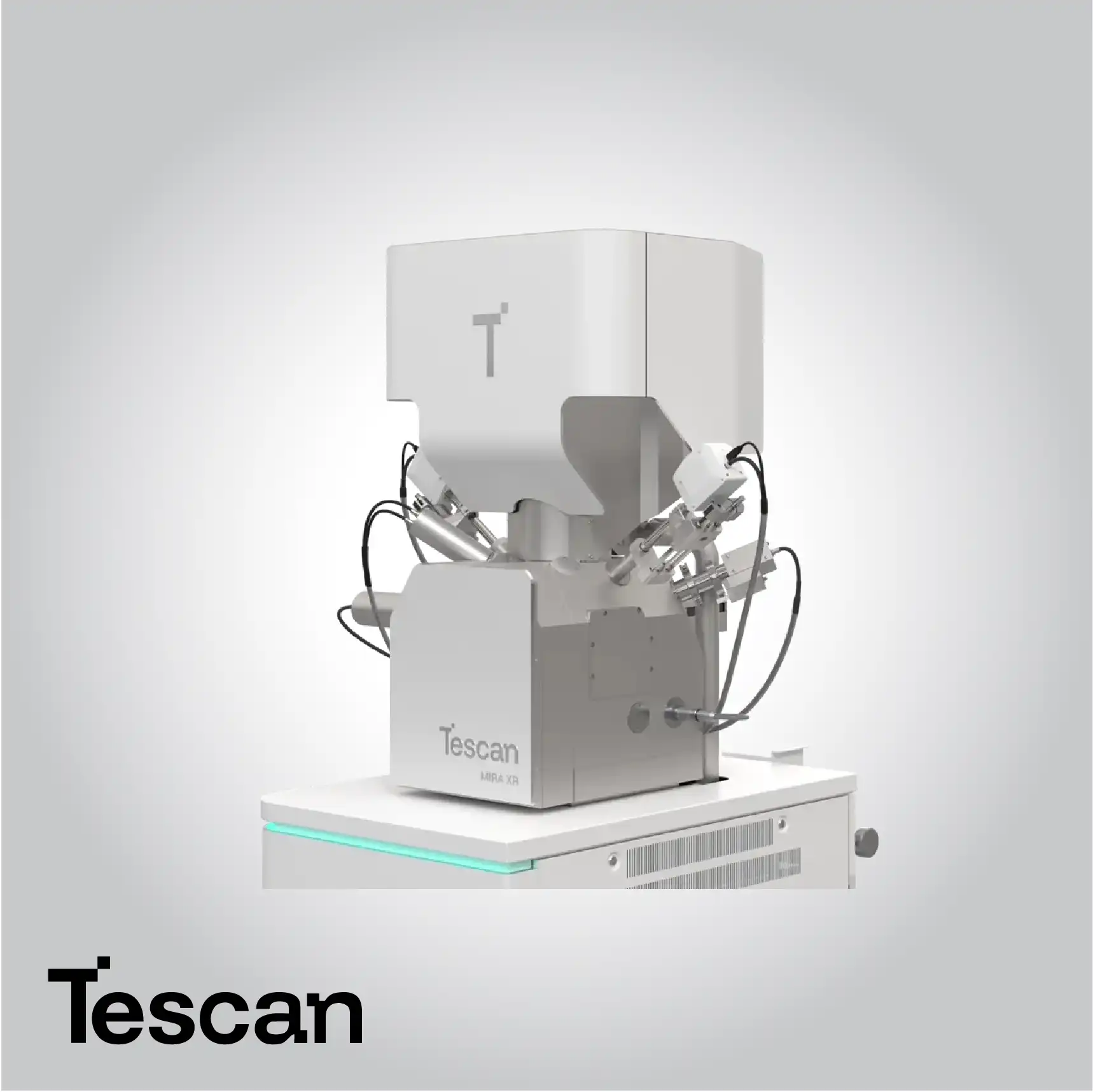
Tescan FIB-SEM systems for life sciences combine high-resolution imaging and precise ion beam milling to reveal the ultrastructural organization of biological specimens in three dimensions. Designed to handle the delicate nature of biological materials, these systems provide a stable, high-vacuum or cryogenic environment that preserves cellular architecture while enabling accurate structural and compositional analysis. The integration of focused ion beam (FIB) and scanning electron microscopy (SEM) technologies allows researchers to perform serial surface imaging, volume reconstruction, and site-specific sectioning with nanometer precision.
Through a synergy of beam stability, advanced detectors, and correlative imaging workflows, Tescan FIB-SEM systems extend the boundaries of life science research. They enable multi-modal 3D reconstruction, targeted sample modification, and high-contrast imaging across a range of biological applications—from cellular analysis and tissue morphology to biomaterial interfaces. The systems’ compatibility with cryogenic workflows preserves hydrated biological samples in their native state, ensuring accurate visualization of intracellular structures and molecular interactions. With precise control over ion beam parameters and minimal sample damage, Tescan FIB-SEM platforms provide scientists with the ability to correlate structure, function, and composition at unprecedented resolution.
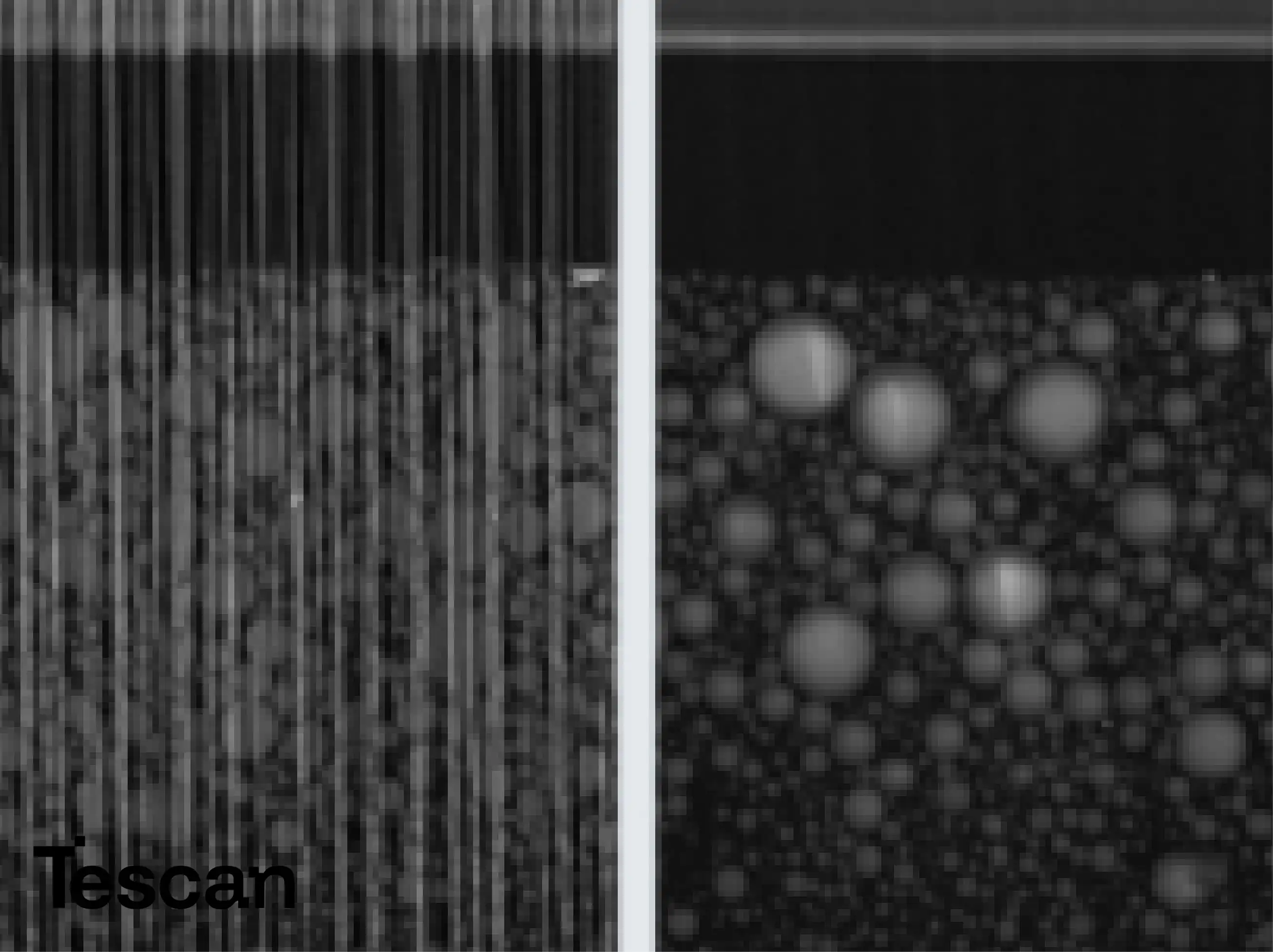
3D Volume Reconstruction for Biological Insight
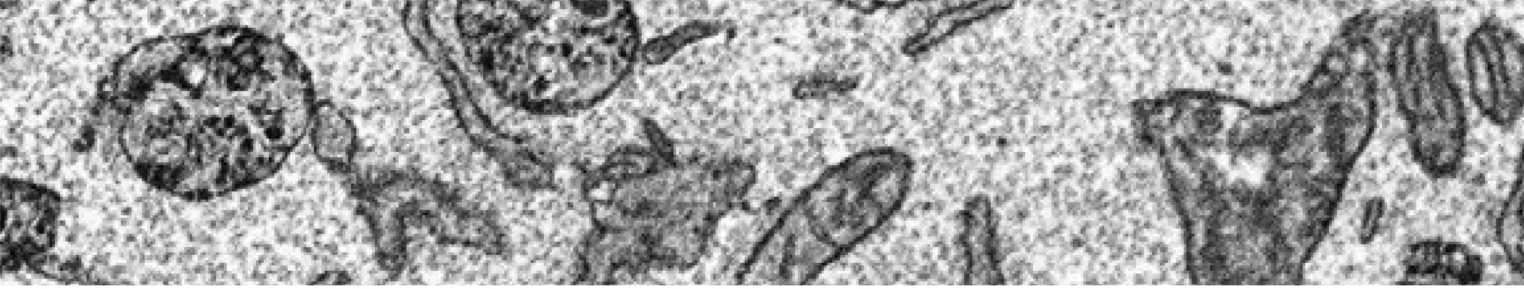
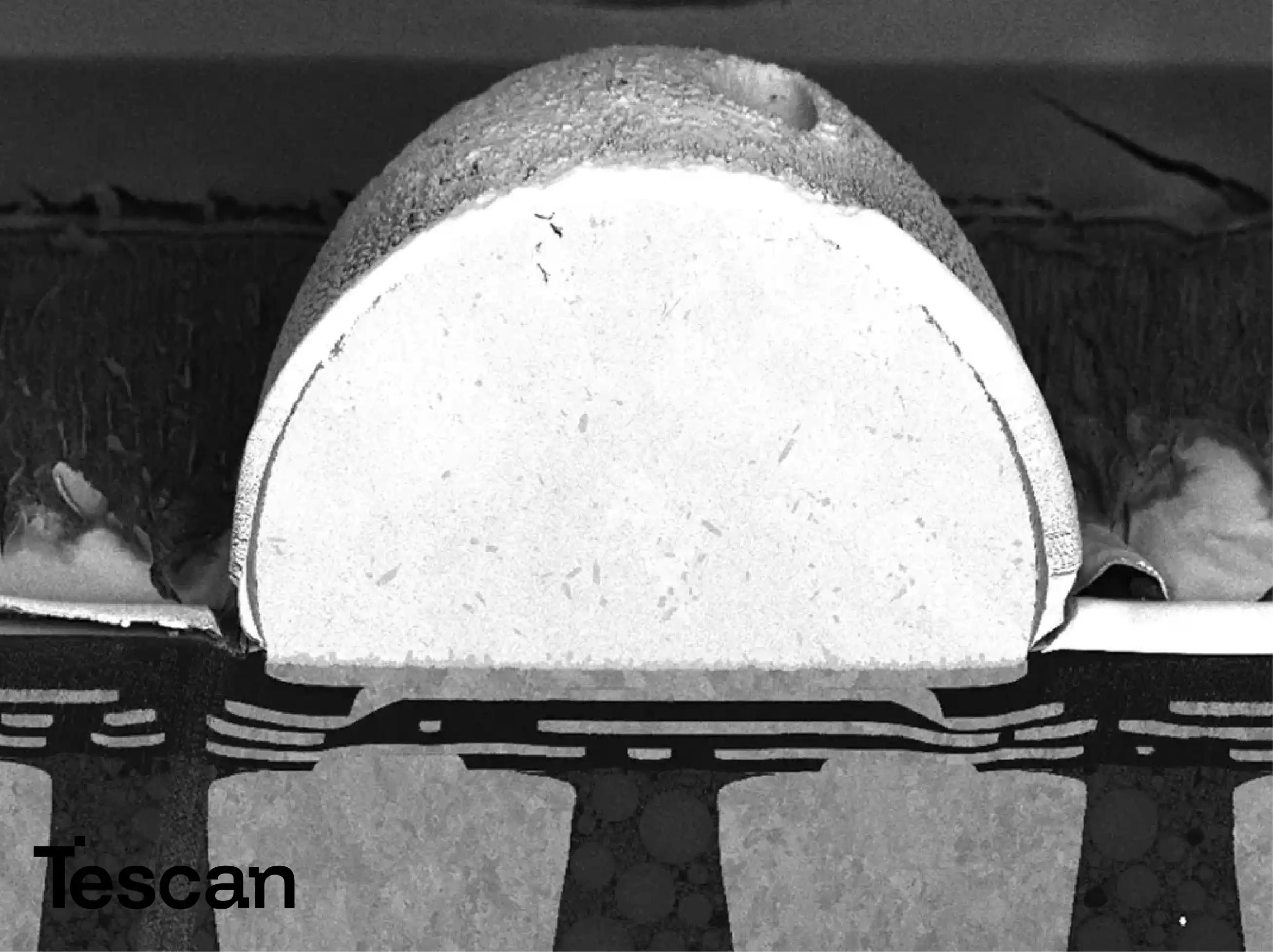
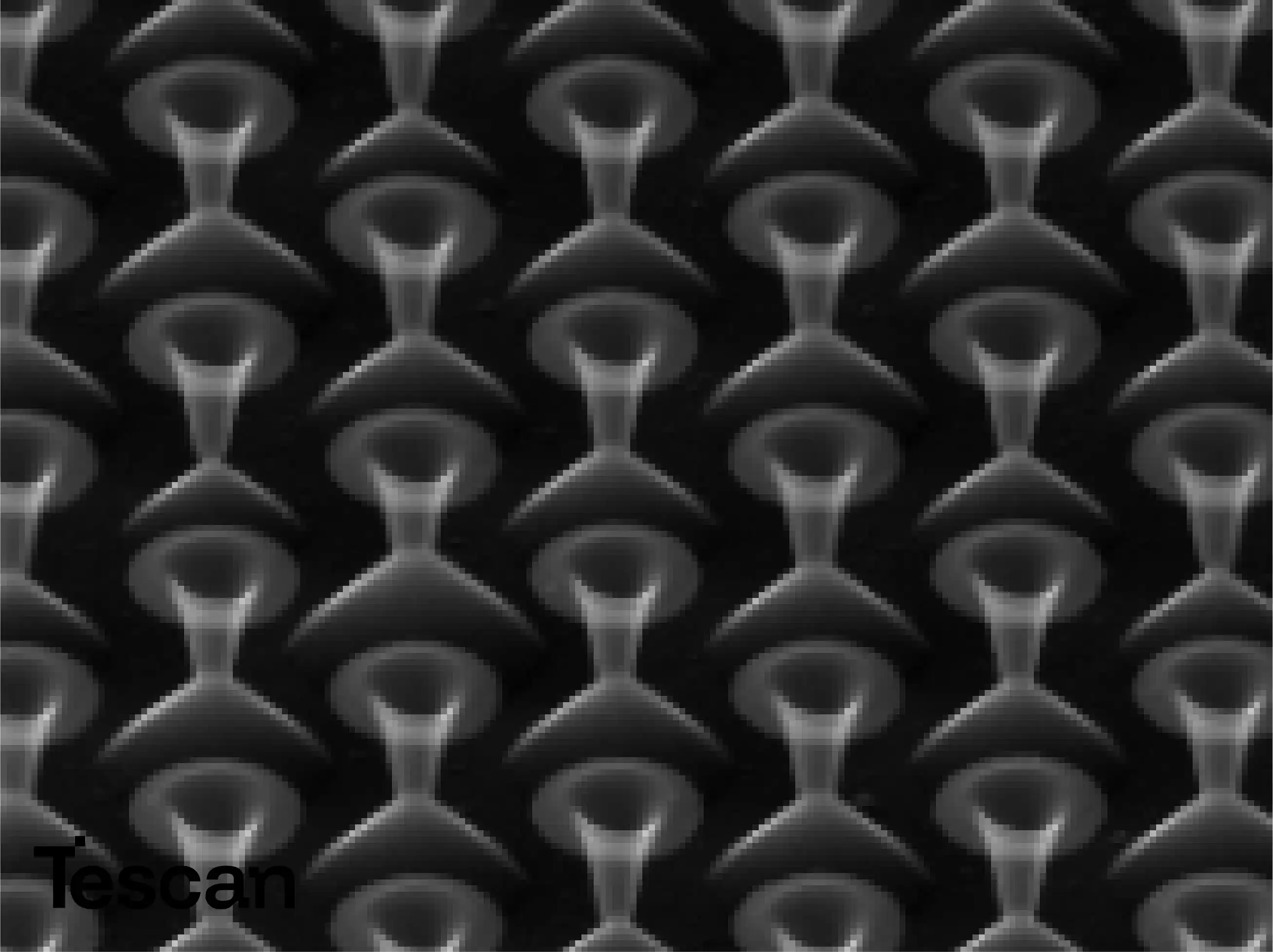
TESCAN FIB-SEM systems enable automated serial sectioning and image acquisition, producing high-resolution 3D datasets that capture the internal organization of cells and tissues. By combining focused ion beam milling with rapid SEM imaging, researchers can reconstruct biological volumes with nanometer-scale detail, allowing for quantitative measurements and spatial mapping of ultrastructural components. This capability supports advanced studies in neuroscience, developmental biology, and biomaterials research.
Cryogenic Imaging for Native Sample Preservation
Cryo-compatible FIB-SEM workflows maintain biological samples in a near-native frozen-hydrated state, minimizing artifacts caused by dehydration or chemical fixation. This enables accurate imaging of lipid membranes, macromolecular assemblies, and cellular organelles. The cryogenic environment ensures structural fidelity, making it ideal for investigating dynamic biological processes and fragile tissues that are otherwise difficult to preserve under conventional vacuum conditions.
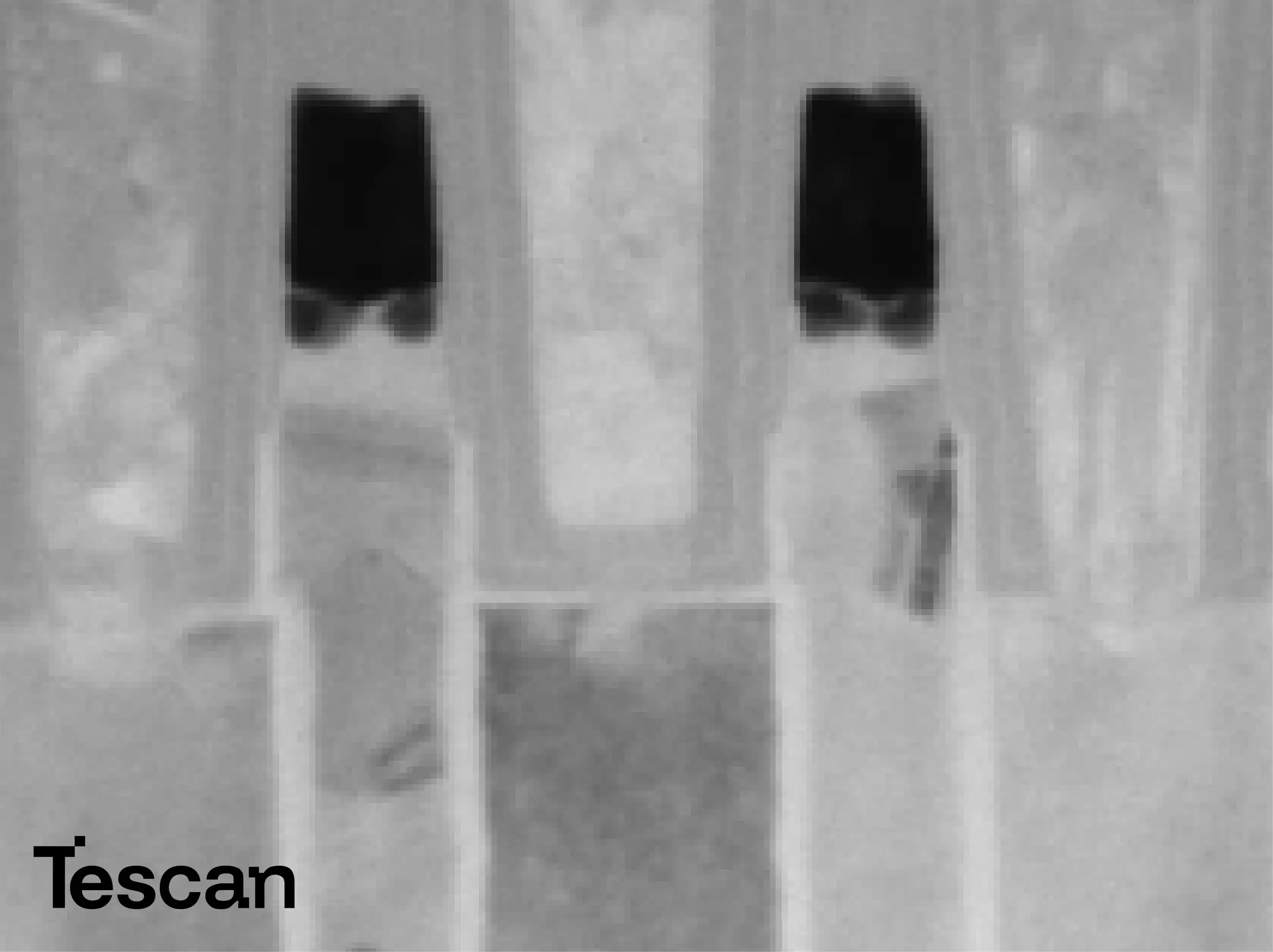
Precise Ion Beam Milling for Serial Sectioning
The focused ion beam provides nanometer-scale precision in material removal, allowing researchers to generate sequential cross-sections through biological samples. This method enables detailed exploration of intracellular and extracellular architectures without mechanical sectioning. Controlled milling ensures smooth layer removal and consistent imaging quality, producing datasets suitable for 3D reconstruction and volumetric analysis of complex biological systems.
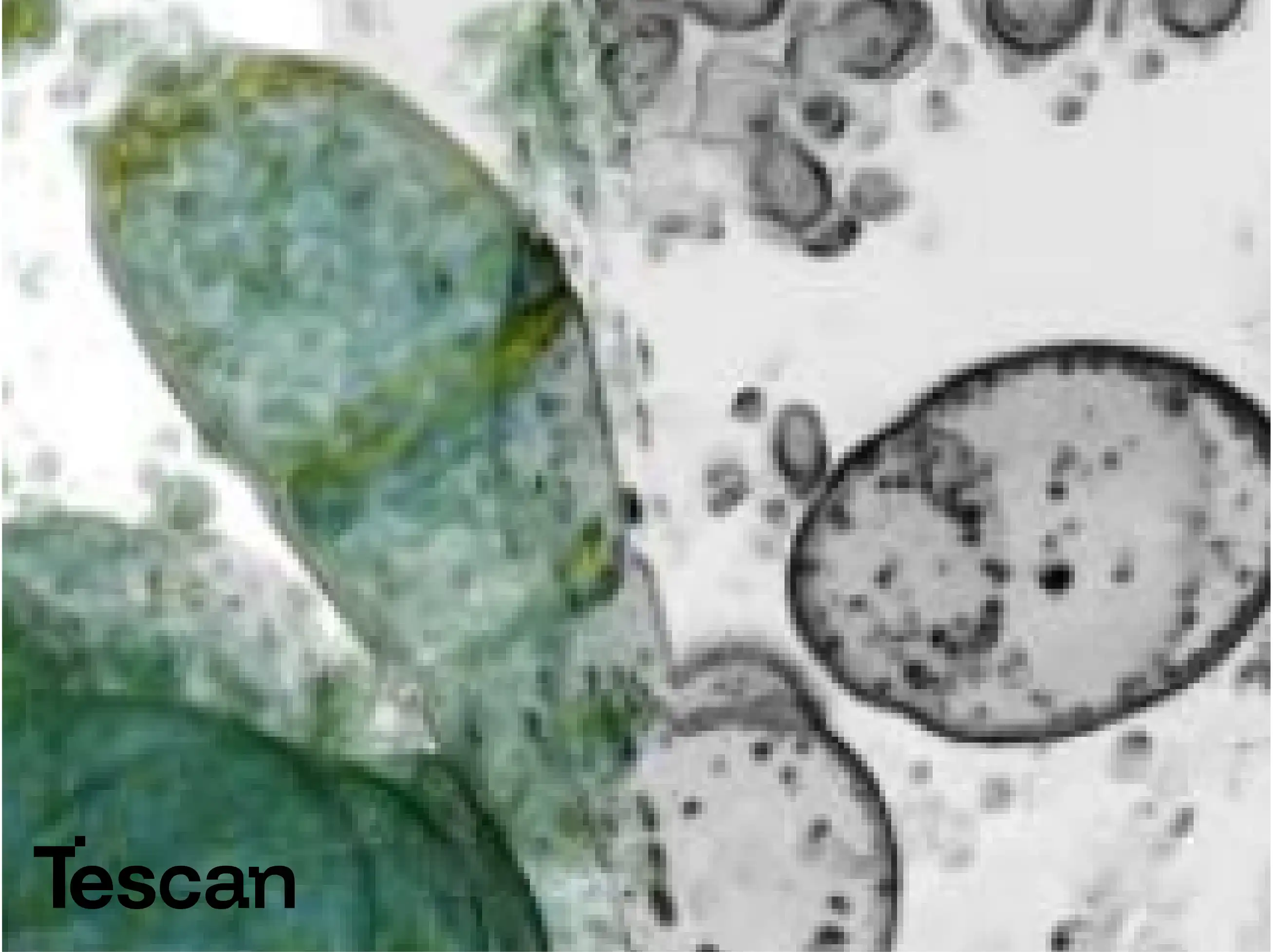
Correlative and Multi-Modal Imaging Workflows
TESCAN FIB-SEM platforms support correlative imaging workflows, integrating data from light microscopy, cryo-fluorescence, and electron microscopy. This allows researchers to localize regions of interest identified in fluorescence imaging and analyze them at nanoscale resolution within the FIB-SEM. By combining complementary imaging modalities, users gain a comprehensive understanding of both the structural and molecular organization of biological samples.
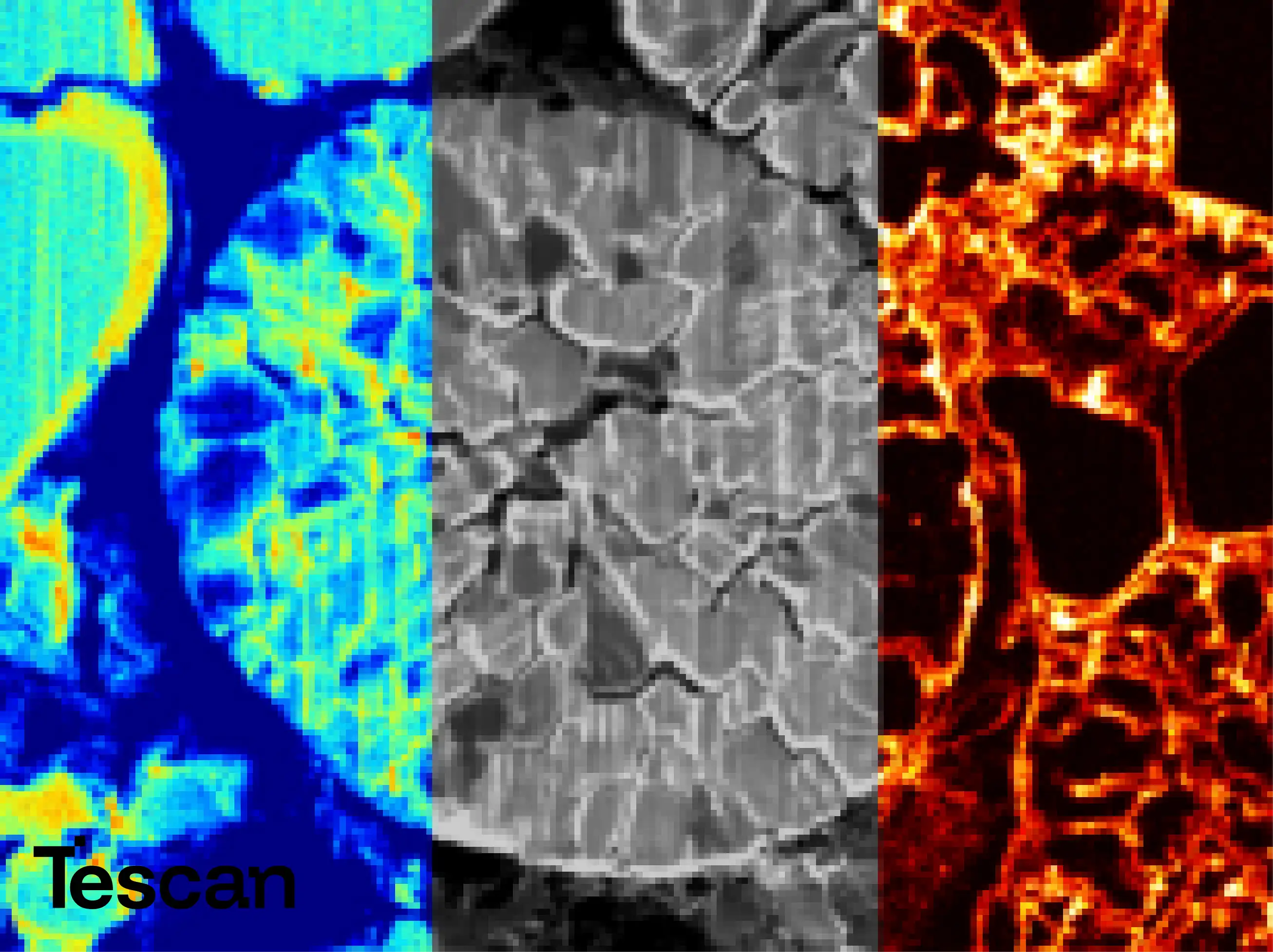
High-Contrast Detection and Imaging Optimization
Equipped with advanced electron and ion detectors, TESCAN FIB-SEM systems deliver high signal-to-noise ratios and optimized contrast for biological specimens. The detection system enhances imaging of low-Z materials and soft biological tissues, ensuring detailed visualization of cellular membranes, nuclei, and vesicular structures. Fine control over imaging parameters such as beam energy and current further improves resolution while minimizing beam-induced damage.
Application Versatility Across Life Science Disciplines
The versatility of TESCAN FIB-SEM systems makes them suitable for a wide range of life science applications, including ultrastructural cell biology, tissue morphology, neuroanatomy, and biomaterial interaction studies. Researchers can investigate the interface between biological tissues and synthetic materials, explore the nanoscale organization of neural circuits, and perform correlative volume analyses across different biological scales—from organelles to entire cell networks.
Advanced Analytical Integration
In addition to imaging, TESCAN FIB-SEM systems support integrated analytical techniques such as energy-dispersive X-ray spectroscopy (EDS) for elemental analysis. This enables compositional studies of biomineralized tissues, implants, or hybrid materials within a biological matrix. Combining imaging and microanalysis within a single instrument provides a holistic understanding of structure–composition relationships in biological systems.
Automation and Data Processing Tools
TESCAN’s advanced software ecosystem automates image acquisition, alignment, and volume rendering, reducing user workload while ensuring consistent data quality. Intelligent pattern recognition and adaptive milling algorithms streamline long-term serial imaging tasks. High-performance visualization tools allow researchers to analyze and interpret 3D datasets efficiently, facilitating quantitative morphometric analysis and publication-ready visual output.
Read more about Tescan products here