Home » Products » Scanning Electron Microscopy » Micro-computed tomography (micro-CT) » Tescan Biological Applications for Life Science
Tescan Biological Applications for Life Science
High-Resolution Micro-CT Imaging for Life Science and Biological Research
Tescan Biological Applications for Life Science
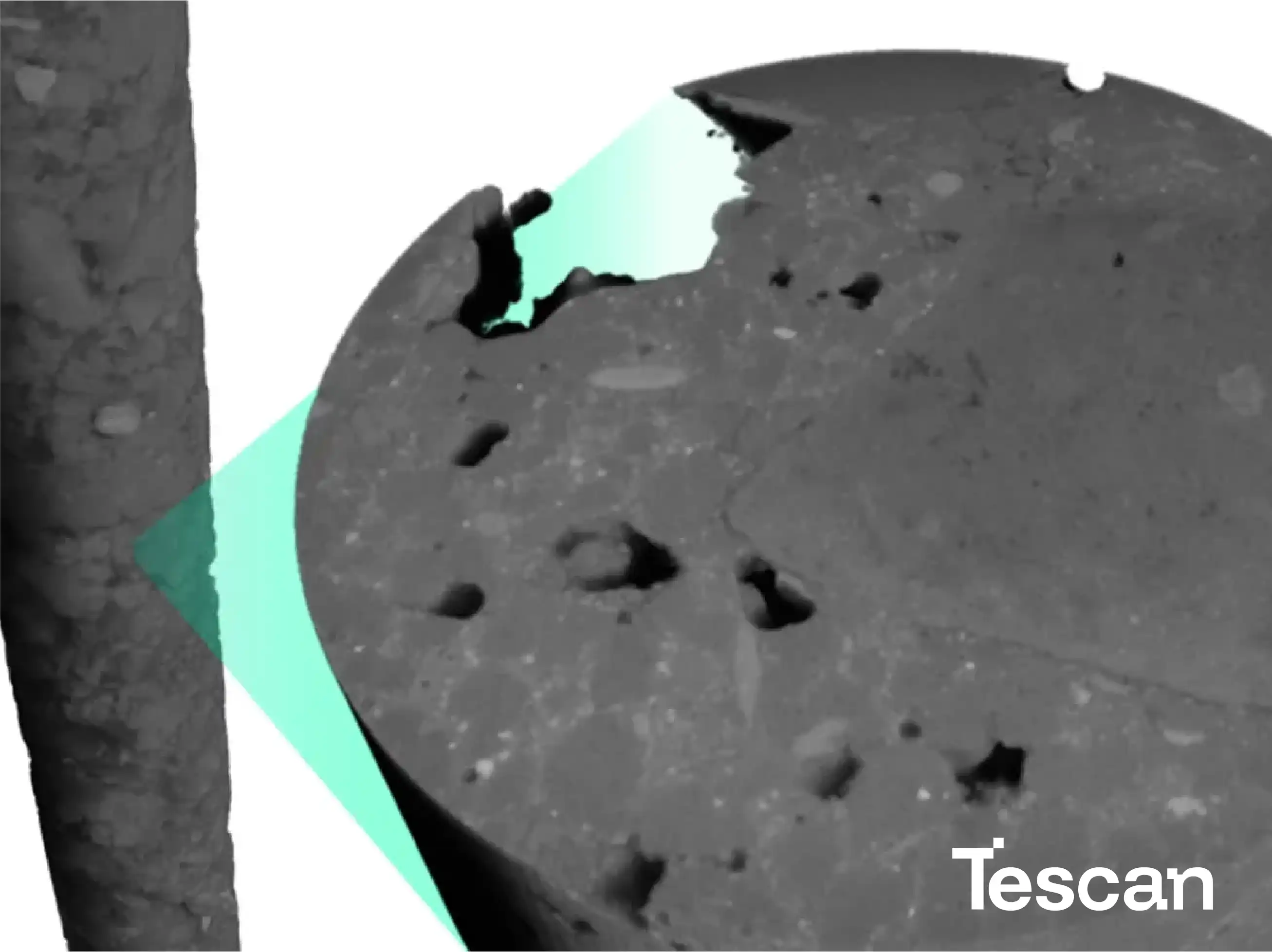
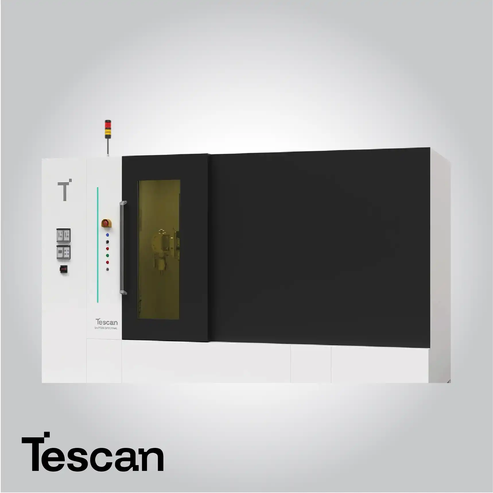
Tescan Micro-CT systems for life science offer non-destructive 3D imaging solutions designed to study biological samples ranging from small tissues to complete organs. These systems preserve the native structure of delicate samples while providing high-resolution volumetric reconstruction. By combining optimized X-ray sources with high-sensitivity detectors and advanced imaging algorithms, researchers can visualize complex internal features such as tissue architecture, vascular networks, and microstructural details at unprecedented clarity.
These platforms support versatile sample types, including hydrated or sensitive tissues, enabling a wide range of biomedical applications. High-resolution Micro-CT imaging facilitates studies in anatomy, pathology, and comparative biology, supporting researchers in understanding structural relationships at multiple scales. Advanced software tools allow volumetric analysis, quantitative measurement of microstructures, and visualization of subtle variations in density and morphology. This combination of precise imaging and analytical flexibility makes Tescan Micro-CT an indispensable tool for modern life science research, from fundamental investigations to translational biomedical studies.
Sample Preservation and Preparation
Tescan Micro-CT systems provide highly optimized conditions to preserve the integrity of delicate biological samples throughout the imaging process. Specialized sample holders and precise positioning mechanisms ensure that tissues, organs, or small organisms remain stable, preventing structural deformation or drying that could compromise image quality. Automated sample handling workflows allow consistent orientation and reproducible positioning, while integrated alignment routines reduce variability between experiments. This ensures that data collected from multiple samples can be directly compared, facilitating longitudinal studies and comprehensive analyses of biological structures.
Volumetric 3D Reconstruction
High-resolution volumetric reconstruction allows researchers to create detailed three-dimensional models of biological samples, revealing tissue layers, organ interiors, and microvascular networks. The interactive visualization tools enable virtual sectioning, enabling examination of internal structures without physically altering the sample. Researchers can explore complex anatomical arrangements, identify regions of interest, and perform precise measurements of structures at various scales. This non-destructive approach preserves the sample for future studies and enables iterative analysis over time.
Quantitative Analysis of Biological Structures
Advanced software tools integrated with Micro-CT systems allow comprehensive quantitative analysis of biological structures. Measurements such as tissue volume, surface area, porosity, density variations, and morphological parameters can be accurately assessed. These quantitative metrics are essential for studies in organ development, tissue engineering, and microstructural pathology, providing researchers with objective and reproducible data that can be correlated with functional, molecular, or experimental variables.
Imaging Hydrated and Soft Tissues
Tescan Micro-CT accommodates hydrated and soft biological tissues without compromising imaging quality. This capability is critical for preserving the natural state of samples, maintaining structural elasticity, and avoiding artifacts introduced by drying or fixation. The system’s high-sensitivity detectors and optimized imaging parameters ensure that even subtle differences in density or texture can be detected, supporting detailed studies of soft tissue architecture, vascularization, and tissue interfaces in their native state.
Comparative Anatomy and Morphology Studies
The high-resolution datasets generated by Tescan Micro-CT enable detailed comparative studies of anatomical structures across different species, experimental conditions, or developmental stages. Researchers can detect subtle variations in organ morphology, tissue organization, and developmental patterns. This capability supports a wide range of biological research, including evolutionary biology, developmental biology, and phenotypic analysis, by providing accurate 3D visualizations that allow meaningful comparisons between samples.
Integration with Multimodal Imaging Techniques
Micro-CT data can be combined with complementary imaging modalities, such as histology, fluorescence microscopy, or MRI, to produce correlative datasets. This integration enables researchers to link structural, functional, and molecular information, creating a more complete understanding of biological systems. Multimodal analysis supports interdisciplinary research by providing cross-scale insights, connecting the microstructural organization observed in Micro-CT with molecular and functional data obtained through other methods.
High-Throughput Imaging and Workflow Automation
Automation in Tescan Micro-CT systems allows multiple biological samples to be scanned efficiently with minimal user intervention. High-throughput workflows enable standardized acquisition of volumetric datasets across large sample cohorts, improving reproducibility and accelerating research timelines. Automated reconstruction, calibration, and analysis routines reduce operator dependency, allowing laboratories to perform large-scale studies such as tissue morphometry, vascular mapping, or organ-level phenotyping with enhanced consistency.
Applications in Biomedical Research and Translational Studies
Tescan Micro-CT supports a wide spectrum of applications in life science research, from basic anatomical and physiological studies to advanced translational investigations. Researchers can study organogenesis, tissue engineering, vascular networks, pathological alterations, and preclinical models of disease. Detailed 3D imaging combined with quantitative analysis provides critical insight into biological structure and function, enabling hypothesis testing, model validation, and the translation of laboratory findings to biomedical applications or therapeutic development.
Read more about Tescan products here


