Home » Products » Scanning Electron Microscopy » Micro-computed tomography (micro-CT) » Tescan Specialized Applications
Tescan Specialized Applications
|
Spectral Micro-CT for Material Discrimination & Multi-Energy Imaging
Tescan Specialized Applications
The Tescan Spectral CT add-on expands the analytical capabilities of Micro-CT imaging by introducing multi-energy acquisition, enabling the differentiation and quantification of materials with similar densities but distinct elemental compositions. This advanced imaging technique provides enhanced contrast and improved segmentation accuracy, allowing researchers to generate detailed compositional maps in three dimensions. Through multi-energy scanning, Spectral CT enhances non-destructive characterization, offering insights into chemical and structural variations that traditional X-ray absorption imaging cannot distinguish.
By integrating advanced detector technology and precise energy calibration, the Spectral CT system captures multiple energy levels within a single scan, producing datasets that reveal subtle compositional gradients and multi-phase structures. These capabilities are particularly beneficial in analyzing composites, geological samples, batteries, and biological specimens where material contrast is limited. The add-on’s compatibility with dynamic and in-situ imaging workflows enables simultaneous observation of chemical and physical transformations.
Multi-Energy X-ray Imaging: Expanding the Analytical Frontier
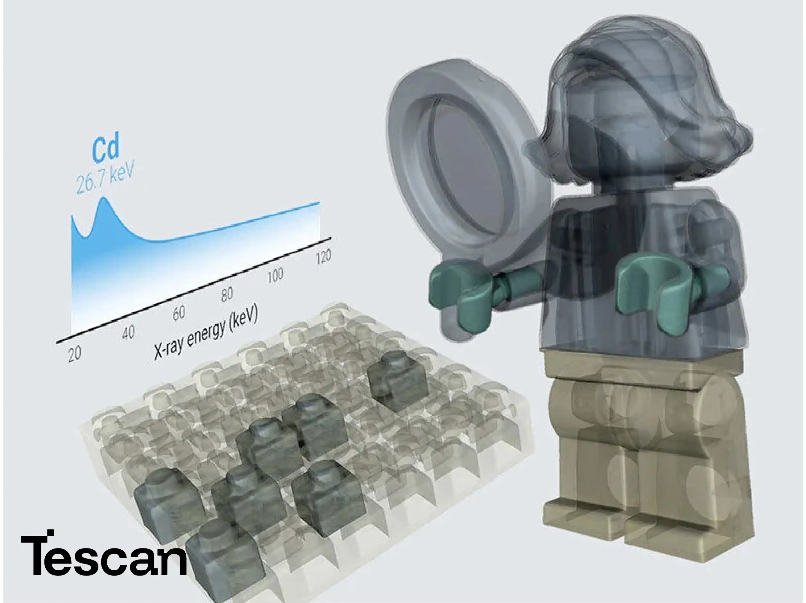
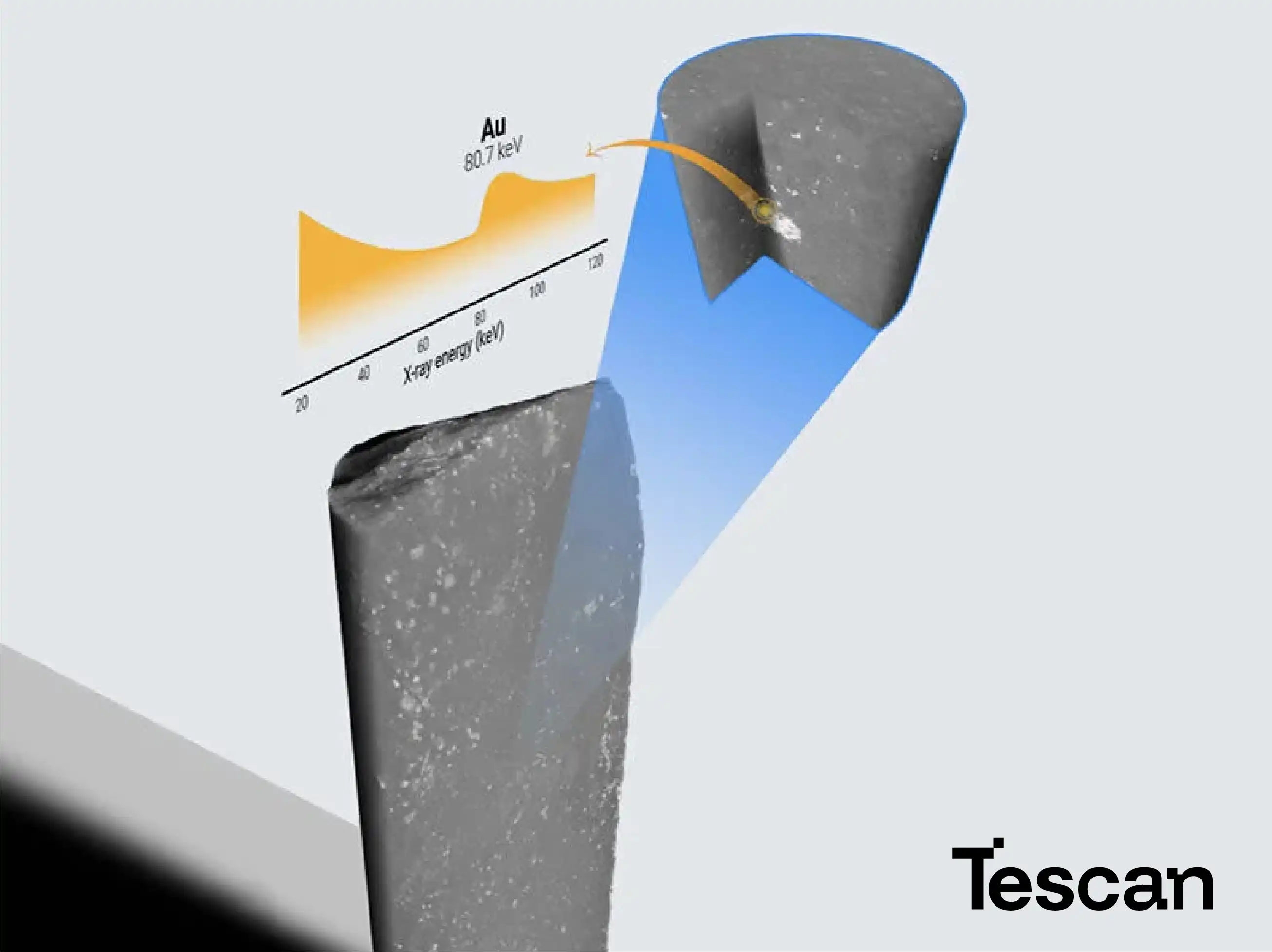
Spectral CT technology introduces a breakthrough in micro-CT imaging by capturing datasets across multiple X-ray energy levels. Unlike conventional single-energy CT, which relies solely on absorption contrast, multi-energy imaging enables direct differentiation of materials based on their energy-dependent attenuation profiles. This allows users to separate elements and compounds that otherwise appear identical in grayscale imaging. By acquiring data at different energy windows and combining them through advanced reconstruction algorithms, Spectral CT delivers a comprehensive visualization of material composition. This approach enhances precision in detecting microstructural variations, supports the identification of multiple materials within a single voxel, and provides a powerful analytical basis for understanding multi-phase or heterogeneous samples.
Enhanced Contrast and Precise Material Segmentation
One of the defining advantages of the Spectral CT add-on is its ability to significantly enhance image contrast and enable precise segmentation of materials with overlapping densities. Traditional CT imaging often struggles to distinguish between phases with similar X-ray absorption characteristics, leading to ambiguity in interpretation. Spectral CT overcomes this limitation by leveraging differences in the energy-dependent attenuation of materials, generating contrast maps that clearly delineate structural boundaries. Researchers can isolate phases, quantify distribution, and perform accurate volumetric analysis of each component. This capability is particularly crucial for composite materials, biominerals, and geological samples, where clear visualization of interfaces and internal structures directly impacts scientific and industrial understanding.
Compositional Mapping and Multi-Phase Quantification
The TESCAN Spectral CT add-on transforms standard micro-CT imaging into a tool for compositional mapping and quantitative phase analysis. By analyzing how materials absorb X-rays across multiple energies, the system creates voxel-by-voxel compositional maps that reveal the internal chemical architecture of a sample. Using spectral decomposition algorithms, each material is assigned a unique spectral signature that can be used to determine its distribution and volume fraction. This quantitative insight enables advanced studies in fields such as battery development, catalyst analysis, and material aging, where understanding the spatial distribution of elements or phases is vital. Researchers can visualize compositional gradients, detect impurities, and assess structural integrity in three dimensions without any sample destruction.
Non-Destructive Elemental Differentiation and Preservation
Spectral CT represents a paradigm shift in non-destructive analytical imaging. It enables researchers to identify and distinguish materials based on elemental composition without physical sectioning, staining, or other invasive preparation. This approach is particularly valuable for delicate biological tissues, rare minerals, and high-value manufactured components where preservation is essential. The system allows precise identification of materials such as polymers, ceramics, and metals that share similar density profiles but differ in atomic composition. Moreover, this non-destructive capability extends the life of valuable specimens, allowing repeat analyses, long-term monitoring, and cross-validation with complementary microscopy or spectroscopy techniques—all while maintaining the integrity of the original sample.
Real-Time Integration with Dynamic and In-Situ Studies
When combined with in-situ stages or dynamic imaging setups, the Spectral CT add-on provides an unparalleled view into the evolution of materials over time. Users can monitor compositional and structural transformations in response to heat, stress, chemical reactions, or fluid flow—all while collecting spectral data that reveals how elemental distributions shift during these processes. This capability is indispensable for studying reactive materials, corrosion behavior, phase transitions, and degradation phenomena. The ability to visualize both the physical and chemical aspects of material change in real-time transforms Spectral CT into a multidimensional analytical platform that bridges structure, composition, and performance.
Advanced Applications in Energy and Battery Research
Spectral CT is particularly transformative in the study of energy storage materials, such as lithium-ion batteries and solid-state electrolytes. It allows visualization of electrodes, current collectors, and separator layers in unprecedented detail—distinguishing between different chemical components that may share similar densities. Researchers can monitor elemental migration, detect local compositional inhomogeneities, and analyze degradation mechanisms over repeated charge-discharge cycles. This level of spectral insight aids in optimizing material composition and architecture for higher energy density and longer battery life. By linking 3D chemical data to electrochemical performance, Spectral CT supports predictive modeling and accelerates the development of next-generation energy materials.
Geological and Mineralogical Material Characterization
In earth and planetary sciences, the TESCAN Spectral CT add-on enhances the ability to discriminate between mineral phases and map complex geological structures in three dimensions. Minerals often exhibit similar densities but differ in atomic number and elemental makeup, making conventional CT insufficient for compositional mapping. Spectral CT captures these differences, providing high-contrast imaging that reveals mineral boundaries, inclusions, and porosity networks. This capability supports quantitative evaluation of ore textures, phase distributions, and mineral alteration pathways. It is also invaluable in understanding diagenetic processes, sedimentary structures, and microfossil compositions, ultimately enabling geoscientists to study natural materials in their undisturbed state.
Intelligent Data Processing and Visualization Ecosystem
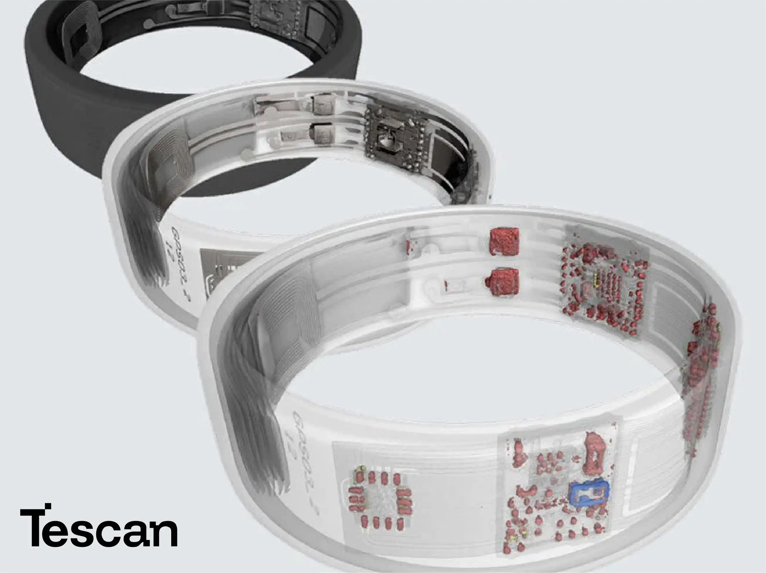
TESCAN’s Spectral CT is complemented by a suite of software tools designed for advanced reconstruction, visualization, and spectral data interpretation. Users can isolate specific energy bands, generate pseudo-colored elemental maps, and perform spectral decomposition to visualize compositional differences with clarity. The software includes modules for quantitative phase analysis, multi-energy histogram plotting, and statistical correlation with material properties. Researchers can also overlay spectral data with microstructural features, building a complete understanding of the sample from both physical and chemical perspectives. This integrated digital workflow transforms complex spectral datasets into actionable knowledge, supporting both academic discovery and industrial innovation.
Read more about Tescan products here