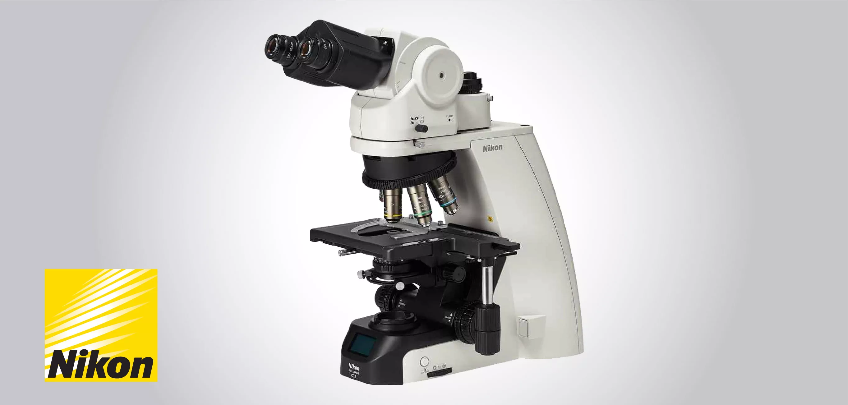
Eclipse Ci-L plus
|
Upright microscope
Nikon announces the new upright microscope Eclipse Ci-L plus
29-09-2021
The new Eclipse Ci-L plus from Nikon Instruments is equipped with Light Intensity Management (LIM) function and LCD which is designed for ease-of-use as well as the health and working styles of people working in research.
With greater comfort and usability, the Ci series brings revolutionary changes to microscopy observation.
The Eclipse Ci-L plus microscope features:
1- Bright and uniform Eco-illumination
• The Eco-illumination is a low-power-consuming, eco-friendly illumination system which generated uniform brightness and decreases the cost and effort of lamp replacement, due to its 60,000 hour, high-luminescent LED. By joining a collimator lens, fly-eye optics and LED illumination, bright and edge-to-edge uniform images can be obtained even at high magnifications. The LED illuminator features low-heat generation and provides the same color temperature at every magnification.
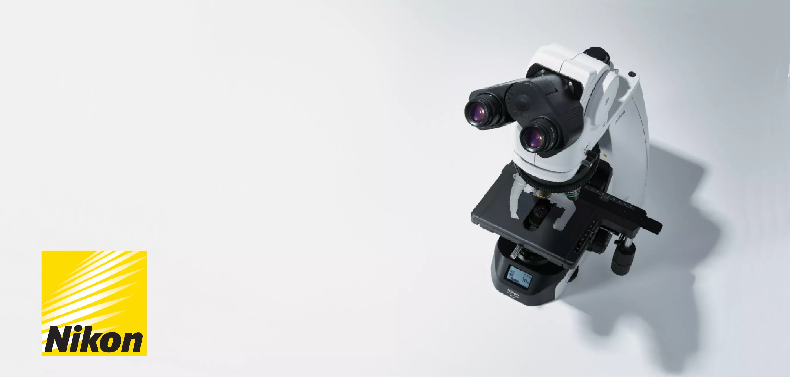
2- Enhanced operational ease
• Observation with a natural posture
Using the ergonomic binocular tube, that features an eyepiece which can be inclined from 10° to 30°and extended up to 40 mm, the microscope can be attuned for natural posture. The eyelevel riser lifts the eyepiece tube in 25 mm increments [up to 100 mm] and can easily be adapted to suit the users with different eye-point heights.
• User-friendly stage operation
With the addition of a nosepiece spacer, the stage height can be lowered 20 mm from the standard position, minimizing strain during frequent specimen change. The stage handle height can also be changed to guarantee a comfortable hand position. The stage height can be locked using the refocusing knob, allowing quick refocusing after specimen changes. The stage is coated with a scratch-resistant high-durability ceramic coating.
3- Easy on the eyes, even during extended use
• Maintains optimal brightness
The light intensity management (LIM) function automatically stores any variations to brightness settings. This helps avoid drastic variations in brightness when switching between changed magnifications during observation, thus helping to mitigate eye strain.
• Keep the same observation posture with automatic brightness adjustment
The LIM function keeps and recalls the optimal brightness level for every objective, minimizing the need to manually adjust the illuminator and change posture every time you switch objective lenses.
4- Effortless image capturing
• One simple click of the image capture button on the microscope base throughout observation allows the Digital Sight camera to capture the specimen image. Camera control software for tablet PC NIS-Elements L is equipped with a scene mode that can automatically set the optimum shooting conditions for each observation method. Since it has a network function, you can share images with a remote PC.
5- Enhanced efficiency during observations
• Status Display for simple confirmation at a glimpse
Easily and quickly check brightness and magnification settings using the display at the base of the microscope deprived of changing your observation posture
• Turning the nosepiece automatically adjusts the scale bar
The scale bar on the PC display changes automatically to match the magnification level, minimizing the need to set the scale manually.
6- Versatile observation techniques
• Phase contrast
High-contrast images with neutral background coloration irrespective of the magnification range can be captured. This observation technique is appropriate for observation of unstained structures.
• Simple polarizing
Perfect for observing bi-refringent samples such as collagen, amyloids and crystals.
• Sensitive color polarizing
Permits Identification of uric acid crystals by changes in the interference color. This technique is perfect for gout and pseudo-gout tests.
• Darkfield
Allows clear observation of blood or minute structures such as flagella. Dry- and oil-type condensers available as well. An expander lens is applied for brighter imaging.
• Epi-fluorescence
Compact epi-fluorescence attachments that exploit noise terminating mechanisms allow weakly fluorescing specimens to be captured with great clarity and brightness. Both CI-FL epi-fluorescence attachment (incorporates up to 4 filter cubes) and D-FL epi-fluorescence attachment (incorporates up to 6 filter cubes) permit easy switching of filter cubes. High-optical-performance objective lenses for epi-fluorescence imaging, including the CFI Plan Apochromat Lambda series and the CFI Plan Fluor series, are available.
.webp)