
Home » Products » Chromatography/Mass Spectrometry » MALDI-Based Instruments and Solutions » Shimadzu MALDI Imaging Solutions
MALDI Imaging Solutions
|
Specialized High-Resolution MALDI Mass Spectrometry Imaging

Shimadzu MALDI Imaging Solutions
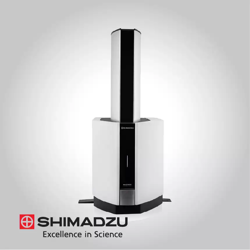
The technical architecture of these solutions is built on a seamless workflow encompassing sample preparation, automated matrix coating, and sophisticated data processing. Systems like the iMScope QT offer a high spatial resolution of up to 5 micrometers, enabled by a 20 kHz high-repetition-rate laser that ensures rapid data acquisition without sacrificing sensitivity.
For laboratories seeking accessible imaging, the benchtop imaging starter kits extend these capabilities to compact linear TOF platforms. By utilizing specialized software for image registration and statistical analysis, these instruments enable researchers to overlay morphological microscopic images with molecular heat maps, revealing the complex chemical signatures hidden within biological tissues.
High-Resolution Atmospheric Pressure MALDI (AP-MALDI)
Shimadzu’s premier imaging systems utilize an Atmospheric Pressure MALDI (AP-MALDI) source, which is technically critical for analyzing volatile or semi-volatile compounds. Unlike vacuum MALDI, the atmospheric environment prevents the sublimation of certain matrix types and preserves the hydration state of biological samples. This interface is optimized for high sensitivity and provides a soft ionization path that minimizes the fragmentation of large biomolecules, making it ideal for the high-definition mapping of small metabolites and large intact proteins in a single experiment.
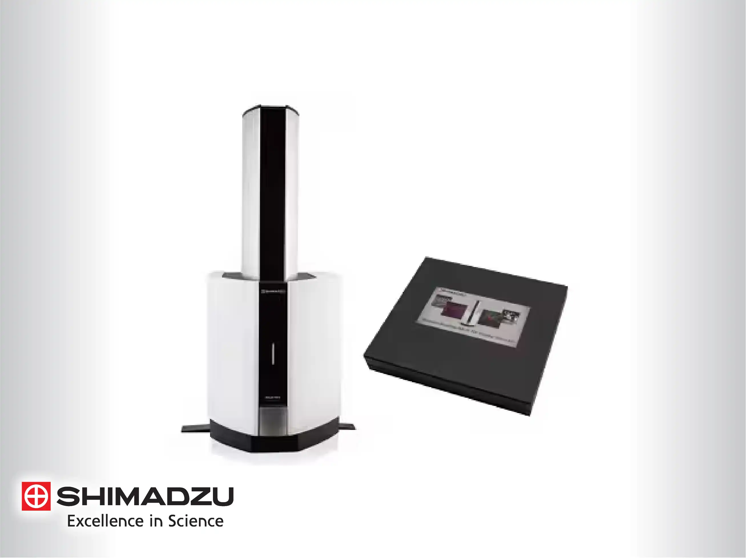
Micro-Resolution Imaging with 5 µm Precision
The imaging mass microscope technology achieves a high spatial resolution of 5 micrometers, allowing for the observation of molecular distributions at the cellular level. Technically, this is achieved through precision-engineered laser optics and micro-stepping sample stages. This level of detail enables researchers to distinguish between adjacent histological structures, such as different layers of the retina or specific regions of a tumor, providing insights into the localized chemical changes associated with various disease states.
High-Speed 20 kHz Laser Technology
For high-throughput imaging, the integration of a 20 kHz solid-state laser is a significant technical milestone. This high repetition rate allows the system to scan large tissue sections at unprecedented speeds while maintaining high pixel density. The laser's high-speed pulsing is synchronized with the detector's data acquisition cycle, ensuring that each pixel in the molecular image represents a complete, high-quality mass spectrum. This speed is essential for large-scale clinical studies where multiple samples must be processed rapidly.
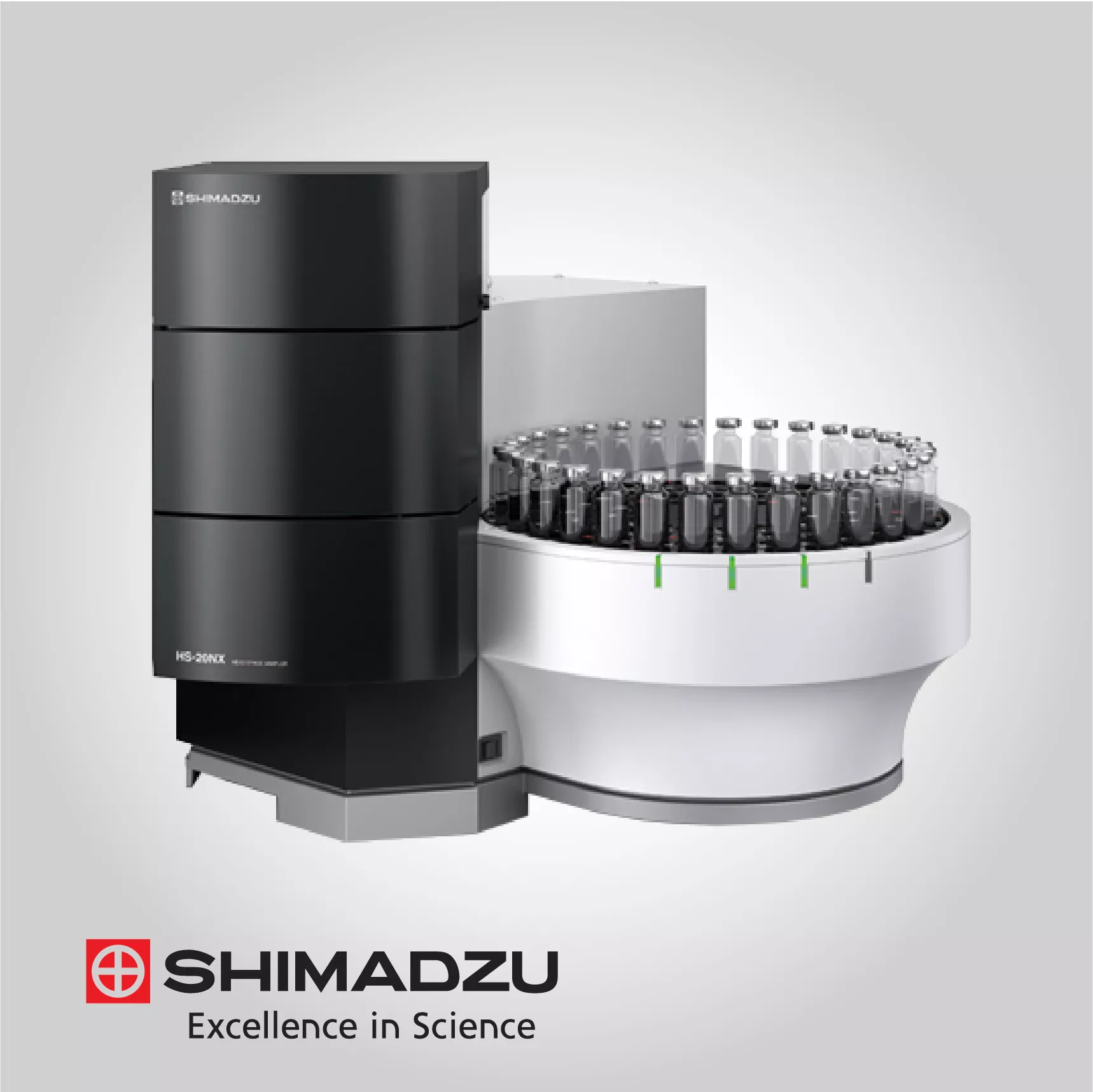
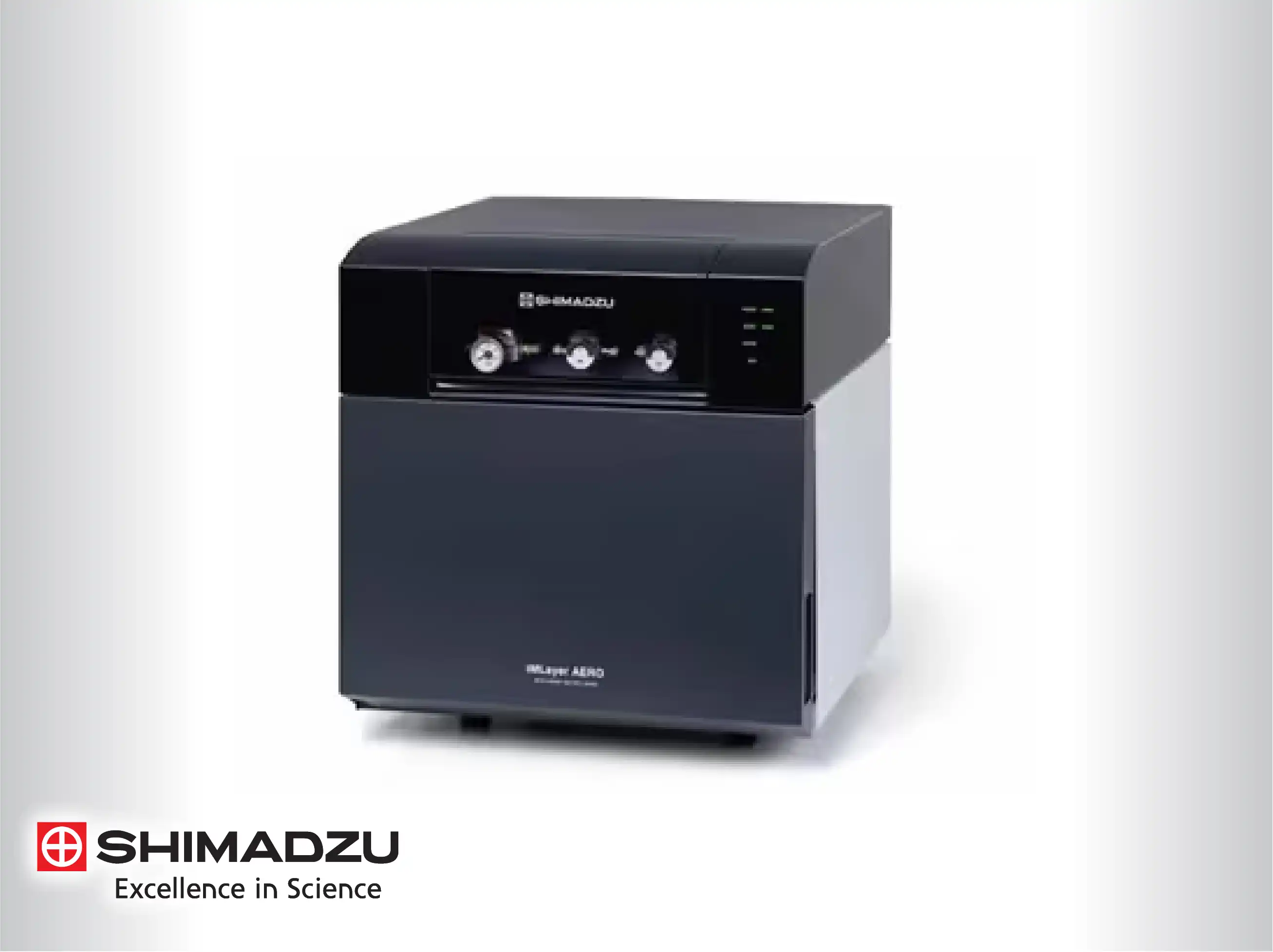
Automated Matrix Coating Systems (iMLayer and Aero)
Sample preparation is technically automated through specialized matrix deposition systems. The iMLayer uses vacuum vapor deposition to create a thin, highly uniform matrix layer with extremely small crystal sizes, which is essential for achieving high spatial resolution below 10 micrometers. Alternatively, the Aero system utilizes automated spraying for sample derivatization and matrix application, providing high reproducibility and flexibility for various tissue types. These automated solutions eliminate operator variability and ensure consistent ionization efficiency across the entire sample surface.
Seamless Fusion of Optical and Mass Spectral Data
The imaging mass microscope technically integrates a full optical microscope within the mass spectrometer housing. This allows for the immediate capture of high-resolution morphological images before MS analysis. The software then co-registers the optical and mass spectral data, enabling users to perform "overlay analysis." By superimposing molecular heat maps onto the optical image, researchers can precisely correlate chemical concentrations with specific anatomical features or pathological changes observed in the tissue.
Versatile Benchtop Imaging Starter Kits
To make imaging technology more accessible, Shimadzu offers benchtop imaging kits designed for linear TOF systems. Technically, these kits include specialized slide adapters (Adaption-mini), ITO-coated glass slides (FlexiVision-mini), and dedicated acquisition software. Despite their compact footprint, these systems provide reliable imaging performance for routine tasks such as lipid profiling or fingermark analysis in forensics. This modular approach allows existing laboratories to upgrade their analytical capabilities without investing in a completely new floor-standing platform.
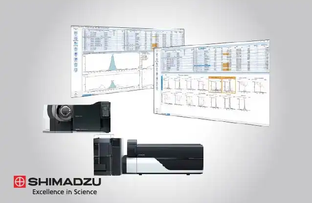
Comprehensive Image Analysis Software (IMAGEREVEAL MS)
The IMAGEREVEAL MS software is a technical cornerstone of the imaging workflow, designed to handle the massive datasets generated during MS imaging. It provides various analysis modes, including principal component analysis (PCA), hierarchical cluster analysis (HCA), and segmentation. These statistical tools allow for the "unbiased" discovery of biomarkers by identifying ions that are spatially correlated with specific regions of interest. The software supports open data formats like imzML, facilitating collaboration and cross-platform data validation.
Quantitative Imaging and Pharmacokinetic Applications
Beyond qualitative mapping, these solutions are technically optimized for quantitative mass spectrometry imaging (qMSI). By incorporating internal standards during the matrix coating process, the systems can determine the absolute concentration of drugs or metabolites in specific tissue regions. This is particularly valuable in drug discovery and development (ADME studies), where understanding the local concentration of an unchanged drug versus its metabolites within a target organ is critical for assessing efficacy and toxicity.
Click here for more information about Shimadzu range of products